
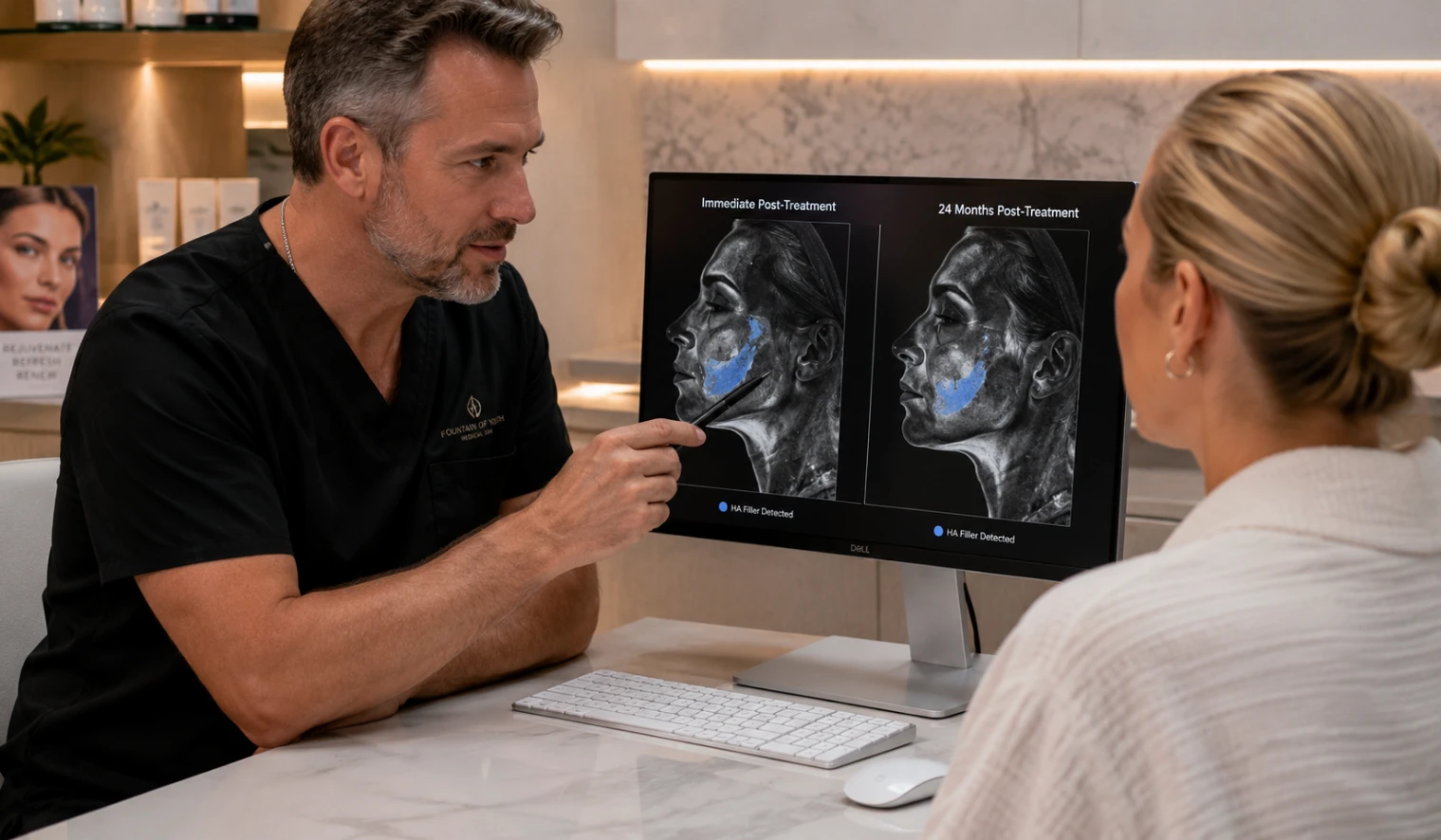
Hyaluronic-acid fillers are usually described as temporary treatments, yet MRI research has complicated that familiar timeline. Detectable material can remain after the visible cosmetic effect has softened or disappeared. People in Fort Myers may interpret a duration estimate as a disappearance date. Current evidence does not support treating those two events as equivalent. A reinjection decision should begin with present anatomy, treatment history, symptoms, and goals.
Long-lasting MRI findings do not mean every filler remains cosmetically useful for years. Imaging signal, visible correction, and a treatment complication answer different clinical questions. Product properties, treatment location, cumulative volume, tissue movement, and individual biology may influence persistence. Small studies cannot establish one lifespan for every patient or product. The practical lesson is careful reassessment, not automatic fear or automatic refilling.
What the Evidence Changes Before Repeat Filler
Visible fading does not prove that every earlier deposit has disappeared. Detectable material does not automatically prove overfilling, migration, or a complication. A useful review connects records, examination findings, photographs, symptoms, and current facial balance. That process may support observation, a smaller adjustment, imaging, referral, or no reinjection.
- Do not treat a product-duration estimate as a guaranteed disappearance date.
- Do not equate an MRI signal with a desirable cosmetic effect.
- Do not schedule repeat volume without identifying a present-day treatment target.
Plan Around Today’s Anatomy
A reinjection review should consider current contour, prior products, treatment dates, injected areas, and new symptoms. Our injectable services begin with an individualized review of present anatomy and history. The objective is proportionate, evidence-aware planning rather than calendar-based maintenance. Some patients may need less product than expected, while others should wait.
Physician-reviewed content supports evidence-aware care and individualized treatment planning for every patient. Published imaging cannot replace an examination by a qualified medical professional. Treatment suitability, product selection, and reinjection timing vary substantially between individual patients.
Compare Established HA Filler Options
Different hyaluronic-acid products are designed for different treatment areas and goals. Review our pages on Juvéderm treatments and Restylane treatments before your consultation. Product selection still depends on anatomy, prior treatment, and the specific concern. A brand name alone cannot determine whether an area needs more volume.
What the MRI Research Shows—and What It Cannot Prove
Recent publications challenge the idea that cross-linked hyaluronic-acid filler always disappears on a predictable schedule. The evidence includes a small MRI series, a single sequential-imaging case, and a clinical illustration involving extensive cumulative treatment. These publications were not designed to create a universal reinjection timetable. They show that long-term persistence can occur under selected circumstances. They do not establish how often persistence occurs across all cosmetic patients.
| Publication | What it reported | What limits the conclusion | Practical meaning |
|---|---|---|---|
| 2024 midface MRI series | All 33 participants showed MRI evidence consistent with residual midface HA after at least two years. | The group was small, histories relied partly on recall, and products were unknown for many participants. | Persistence is possible, but the study does not estimate prevalence in every filler patient. |
| 2025 sequential MRI case | Filler remained visible in the upper lip and cheek region five years after treatment. | The patient had facial surgery and radiation treatment, which may have affected tissue movement. | One unusual case cannot predict longevity for a typical cosmetic patient. |
| 2026 clinical illustration | A heavily treated patient developed progressive fullness and firmness after repeated multiregional filler sessions. | Records were incomplete, cumulative volume was estimated, and the report described one patient. | Cumulative exposure deserves review, but the report remains hypothesis-generating. |
The 33-patient midface series
The 2024 publication reported MRI findings from 33 patients who denied midface injections for at least two years. Every participant showed MRI signal that investigators interpreted as residual hyaluronic-acid filler. Twenty-one reported no midface treatment for two to five years, while twelve reported longer intervals. One patient reported receiving no additional midface injections for more than 15 years. The study found varied signal amounts and did not identify one clearly exceptional product.
The result is clinically interesting, but it should not be converted into a population-wide percentage. Twenty-four participants were recruited without symptoms, while nine underwent imaging because of edema concerns. Treatment histories depended partly on patient recall, and the product was unknown for seventeen participants. Injected volumes, treatment techniques, anatomical locations, and cumulative treatment patterns varied considerably. Larger prospective studies would be needed to estimate typical persistence reliably.
The five-year sequential MRI case
The 2025 case report followed one patient through sequential MRI examinations. Imaging showed persistent material in the zygomatic region and upper lip five years after treatment. The authors reported no evidence of filler migration during that individual case. Facial surgery and radiation therapy may have reduced tissue movement and influenced persistence. Those factors make direct comparisons with routine cosmetic patients unreliable.
The 2026 cumulative-exposure illustration
The 2026 clinical illustration described progressive fullness and focal firmness after repeated treatments spanning roughly two decades. The authors estimated cumulative exposure above 30 milliliters, although precise records were unavailable. Imaging had been publicly disclosed rather than independently collected for that publication. The report proposed layering, compartmentalization, technique, and limited enzyme access as possible contributors. Its conclusions remain cautious because one illustrative case cannot establish a threshold.
Three Questions That Must Stay Separate
The research becomes confusing when persistence, performance, and complications are treated as one issue. Each term describes a different question and requires different evidence. Separating them prevents an imaging result from becoming an automatic treatment decision. It also prevents visible fading from becoming proof that an area is empty. A careful reinjection consultation should answer each of these questions independently.
Is material still detectable?
MRI can show a high-water signal compatible with hyaluronic-acid filler in soft tissue. A positive finding means a water-rich signal remains in the imaged area. It does not directly measure the amount of intact gel still present. An MRI imaging review explains that scans may reflect injected HA, bound water, and water-rich tissue changes. Imaging findings therefore require clinical history, examination, and qualified radiologic interpretation.
Is the cosmetic correction still visible?
Visible improvement depends on more than the physical presence of a deposit. Facial aging continues as fat compartments, skin, bone support, and movement change. A small residual signal may contribute little to current contour. Subtle support may remain useful after the dramatic early result has softened. Photographs, symmetry, movement, palpation, and present goals answer this question more directly.
Has filler migrated or caused a complication?
Migration means material has moved away from the intended treatment site. Persistence in an expected tissue plane does not prove migration. Puffiness can reflect product placement, hydrophilic swelling, inflammation, tissue changes, or another medical issue. Firmness, tenderness, discoloration, progressive swelling, or new asymmetry deserves professional assessment. Social media photographs cannot establish the cause of an individual contour change.
The REST Review Before Reinjection
REST is a practical decision framework created to organize a repeat-filler consultation. It is not a validated clinical guideline or a substitute for professional judgment. The four steps connect historical information with current findings before additional volume is considered. Each framework step can materially change the eventual treatment plan and endpoint. Missing treatment information should increase clinical caution rather than encourage convenient assumptions.
| Step | Question | Useful evidence | Possible outcome |
|---|---|---|---|
| R — Records | What product, amount, location, date, and technique were previously documented? | Treatment summaries, product labels, receipts, injector notes, and dated photographs. | Proceed with clearer context or pause while important records are requested. |
| E — Examination | What does the area look and feel like today? | Contour, symmetry, movement, palpation, swelling, tenderness, firmness, and skin changes. | Treat a defined finding, observe, investigate, or refer when appropriate. |
| S — Signal | Would imaging answer a specific unresolved clinical question? | Disagreement between history and examination, uncertain location, or unexplained symptoms. | Discuss ultrasound, MRI, another evaluation, or no imaging. |
| T — Treatment threshold | What current finding justifies additional product today? | Defined goal, remaining correction, facial balance, alternatives, and conservative endpoint. | Use less product, select another approach, wait, or decline reinjection. |
Why a Filler Timeline Remains an Estimate
Cross-linked hyaluronic-acid gels are designed to resist rapid breakdown after injection. Products differ in concentration, cross-linking, cohesiveness, particle structure, and swelling behavior. Treatment depth and anatomical location influence how a deposit behaves. Highly mobile areas may not behave like deeper, less mobile compartments. A duration estimate describes expected cosmetic performance, not guaranteed molecular disappearance.
Cumulative filler exposure makes simple treatment timelines considerably less reliable over time. One treatment creates a different history from repeated sessions across several compartments. Earlier deposits may overlap with later deposits or remain clinically silent. Incomplete records can obscure how much product has accumulated over time. That uncertainty supports conservative decisions when present anatomy already looks adequately supported.
The FDA explains that duration depends on filler material and injection location. The agency states that repeated long-term use has not been evaluated in controlled clinical studies. FDA guidance also recommends discussing product labeling, injection sites, provider training, and procedure risks. Those points matter even when earlier treatment caused no obvious problem. A routine anniversary should never replace informed consent and current assessment.
When Imaging May Add Useful Information
MRI is not automatically necessary before every consultation involving previous facial filler. A reliable history and normal examination may answer the practical question without a scan. Imaging becomes more relevant when the history and current findings do not align. Uncertain product location, unexplained swelling, firmness, or contour changes can strengthen the discussion. The selected modality should match the clinical question rather than patient anxiety alone.
MRI provides strong soft-tissue contrast and can show water-rich signal across deeper facial planes. It may be useful when a broader anatomical overview is needed. High-frequency ultrasound can provide real-time information about selected deposits and nearby structures. A 2025 ultrasound review found support for filler detection, vascular mapping, and complication assessment. Operator experience and the clinical question influence the value of either test.
| Question | Examination and records | Ultrasound discussion | MRI discussion |
|---|---|---|---|
| Is routine reinjection appropriate? | Usually the starting point for present contour, symptoms, and documented history. | May be unnecessary when history and examination are clear. | Usually unnecessary for straightforward maintenance planning. |
| Where is a selected deposit? | May identify a likely location but cannot map every deposit. | Can provide real-time localization in experienced hands. | Can provide broader anatomical detail across deeper planes. |
| Why is swelling or firmness present? | Essential for identifying timing, tenderness, inflammation, and other warning signs. | May help assess selected deposits, nodules, and nearby blood flow. | May help when deeper or diffuse findings remain unclear. |
| Does imaging prove treatment is needed? | No single examination finding decides treatment without context. | No. Findings must be connected with symptoms and treatment goals. | No. Detectable signal does not establish cosmetic necessity. |
What Different Findings May Mean
The same visible or physical concern can reflect several different clinical situations. A visible change should be interpreted alongside symptoms, examination, photographs, and treatment records. The comparison below organizes reasonable discussion points without diagnosing a patient. No row creates an automatic instruction to inject, dissolve, or image. Individual findings require professional evaluation before treatment decisions are made.
| Finding | What it may indicate | Reasonable discussion |
|---|---|---|
| Visible effect has faded | The original correction softened, although residual material may remain. | Compare present anatomy with photographs before adding volume. |
| Material is detectable on imaging | Water-rich signal remains within the scanned area. | Match location and signal with history, symptoms, and examination. |
| Area looks full but feels normal | Natural anatomy, remaining support, or accumulated product may contribute. | Consider waiting or using less product than a routine schedule suggests. |
| Persistent swelling or firmness | Placement, inflammation, retained material, or another condition may require assessment. | Discuss focused evaluation and whether imaging or referral would help. |
| Contour appears outside the treated area | Migration is possible, but appearance alone cannot confirm it. | Review records, examine the area, and image when clinically useful. |
What to Bring to a Reinjection Consultation
Accurate treatment records become increasingly valuable after several filler treatment cycles. Bring product names, treatment dates, injected areas, approximate amounts, and injector information when available. Product labels, receipts, portal summaries, and photographs can reconstruct an incomplete history. Records from different medical offices may reveal overlapping or cumulative treatment patterns. Clear documentation reduces the need to guess about earlier volume.
Photographs should include comparable angles, lighting, expression, and timing whenever possible. Images can help separate normal aging from fading correction or gradual accumulation. Current symptoms should be described with onset, duration, triggers, and progression. Mention prior dissolving treatments, facial procedures, dental work, infections, or inflammatory episodes. Those details can change the interpretation of swelling or firmness.
Questions worth asking
Ask what present-day finding supports treatment rather than relying on elapsed time. Ask how much correction appears to remain and whether less product could meet the goal. Ask which specific findings would support waiting, imaging, further evaluation, or referral. Ask which alternatives could address the concern without adding volume. A clear plan should define both the target and the stopping point.
Before Adding More Volume
Residual filler does not automatically rule out treatment, but it changes what should be assessed. A consultation through our medical aesthetics service can review present anatomy, photographs, treatment history, and symptoms. The proposed amount should address a current finding rather than a routine anniversary. Unexplained facial changes may require focused evaluation before any elective injection proceeds.
When Routine Reinjection Should Wait
Elective treatment should pause when the area has unexplained swelling, firmness, tenderness, discoloration, or progressive asymmetry. A recent infection, active skin inflammation, or unclear prior product can require further assessment. The purpose of waiting is to identify the problem before changing the anatomy again. Imaging may help in selected cases, but examination remains essential. A cosmetic appointment should not substitute for urgent medical care.
The FDA advises immediate medical attention for unusual pain, vision changes, or possible stroke symptoms. White, gray, or blue skin during or shortly after injection also requires urgent attention. Those symptoms are different from long-term MRI persistence and require urgent evaluation. Delayed concerns still deserve timely professional assessment when they worsen or remain unexplained. Patients should describe exactly when symptoms began and how they changed. Clear timing helps separate routine post-treatment effects from more concerning patterns.
A Better Endpoint Than Chasing a Date
The emerging research supports more careful timing, not fear of all previous filler. Many patients have uncomplicated results, and detectable material may remain without causing a visible problem. The concern becomes practical when contour, symptoms, records, and repeated treatment history do not align. A structured review can identify whether the next step is observation, documentation, imaging, treatment, or referral. No calendar or standard maintenance interval can make that distinction reliably by itself.
Repeat treatment should have a defined reason and a conservative endpoint. Remaining correction, cumulative history, facial proportions, and patient goals matter more than a generic interval. Patients gain useful control by keeping records and asking what supports treatment today. A clinician should explain the target, proposed amount, alternatives, and reasons for waiting. That discussion turns longevity research into better decision-making rather than unnecessary anxiety.
Fountain of Youth in Fort Myers, Florida, reviews filler longevity research within the context of individual treatment planning. Patients can bring earlier records and photographs for comparison during a consultation. The discussion may support treatment, a smaller adjustment, observation, imaging, or referral. Published research cannot determine treatment suitability without an individualized medical examination. Questions about a consultation can be discussed by calling 239-355-3294. Our team can explain scheduling and the records worth bringing.
Who May Benefit From a More Detailed Review?
Repeat filler planning becomes more complex when appearance and treatment history do not align. A focused assessment can identify which missing details matter before another injection. The review should become more deliberate when several years or treatment sites are involved. Persistent symptoms should receive greater weight than any routine maintenance schedule. Several common decision points deserve closer review before elective reinjection:
- You received filler over several years, but product names, amounts, or injection sites remain uncertain.
- The visible effect softened, yet the area still appears supported or fuller than expected.
- You notice persistent swelling, firmness, tenderness, new asymmetry, or unexplained contour changes.
The appropriate next step may be observation, conservative treatment, imaging, or referral. No single evaluation pathway fits every patient, treatment history, or anatomical area. The decision should follow current examination findings and the available treatment records. Missing information should be acknowledged rather than replaced with assumptions. A more detailed review can prevent unnecessary volume and poorly targeted treatment.
Frequently Asked Questions
Does MRI prove that my filler is still working?
No, MRI does not prove that earlier filler remains cosmetically effective or desirable. MRI can show signal compatible with residual water-rich material, but it cannot measure whether the original correction remains desirable. Photographs, examination, facial movement, and current goals provide different information. A scan may detect material that contributes little to visible contour. It may also show signal that requires interpretation alongside tissue changes. Imaging findings should never be treated as a satisfaction score.
Should everyone get imaging before more filler?
No routine clinical rule requires imaging before every repeat filler treatment. Clear records, a normal examination, and an understandable cosmetic change may make imaging unnecessary. Uncertain history, unexplained swelling, firmness, or asymmetry may justify discussing it. The test should answer a defined question that affects management. Imaging without a clinical question may add cost without improving the decision.
Does long-lasting filler always mean migration?
No, long-lasting filler does not automatically establish migration or another complication. Persistence describes material remaining detectable, while migration describes movement away from the intended location. Published MRI reports have documented long-lasting filler without observed migration. A clinician must compare the suspected location with treatment history and current anatomy. Puffiness or asymmetry can have several possible explanations requiring individual assessment. Appearance alone cannot reliably confirm filler migration or identify its cause.
Can I still receive more filler after previous treatment?
Possibly, but timing should depend on current anatomy and the reason for treatment. Residual material does not automatically prevent carefully selected repeat filler treatment. A faded cosmetic result does not automatically require another full syringe. A conservative assessment may support less product, observation, imaging, or another approach. The treatment target should be defined before the amount is selected.
Continue With a More Informed Consultation
Review our guide to navigating injectables and our medical sourcing standards before deciding what to ask. These resources explain how evidence, treatment goals, and clinical context should shape planning. Bring prior treatment records and comparable photographs whenever they remain available. Describe every unexplained contour change before any elective filler treatment begins. A clearer treatment history supports a safer and more proportionate clinical decision.
